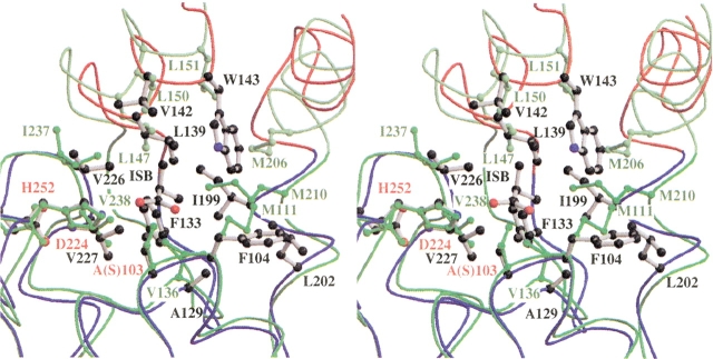
Fig. 7.
Superimpositioning of CumD and RHA1 BphD at the D-part. Viewed from a similar direction as in Fig. 3 ▶. Backbone traces are colored as in Fig. 3 ▶. The catalytic residues and the residues involved in the formation of the D-part of CumD are shown as a ball-and-stick model and are labeled in red and black, respectively. The residues of RHA1 BphD are shown and labeled in green.

