Fig. 5.

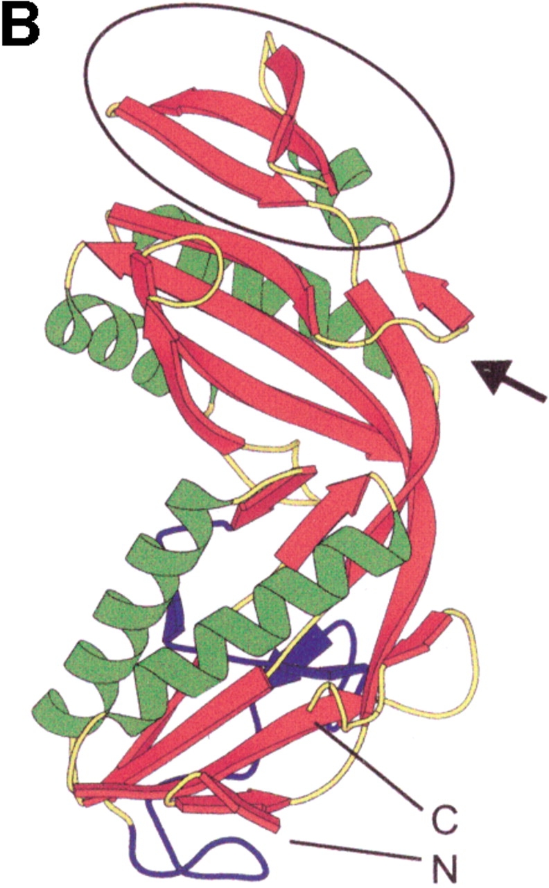
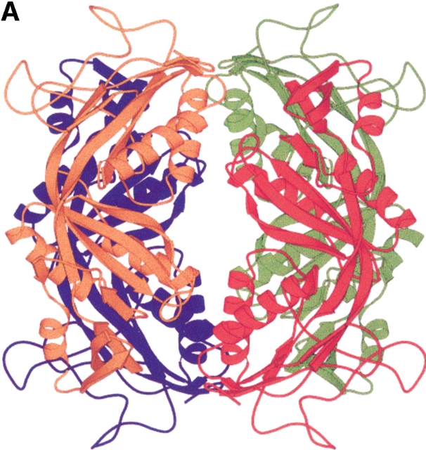
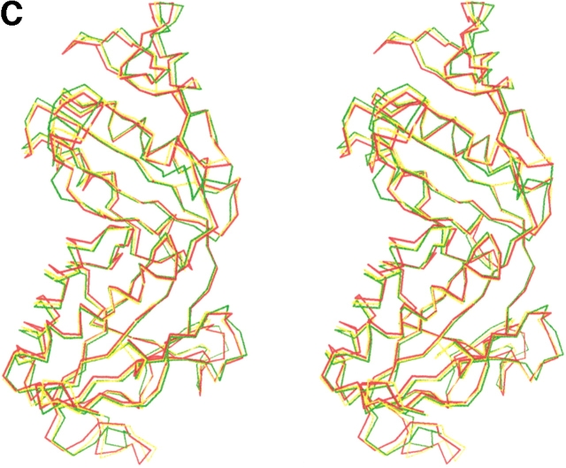
Structure of the formyltransferase. (A) The tetramer presented as a Ribbon diagram indicates a particularly extended contact region between subunits 1 (red) and 2 (green) and the equivalent subunits 3 (blue) and 4 (orange). (B) The Ribbon diagram of the monomer visualizes the location of the insertion region (blue), the meander region (black circle), and the loop between strands 6 and 7 (black arrow). (C) The stereo Cα-plot of the superimposed monomers of the enzymes from M. barkeri (red), A. fulgidus (yellow), and M. kandleri (green) documents their similar fold, in particular, in the core regions of the two lobes. This figure was generated using the program MOLSCRIPT (Kraulis 1991).


