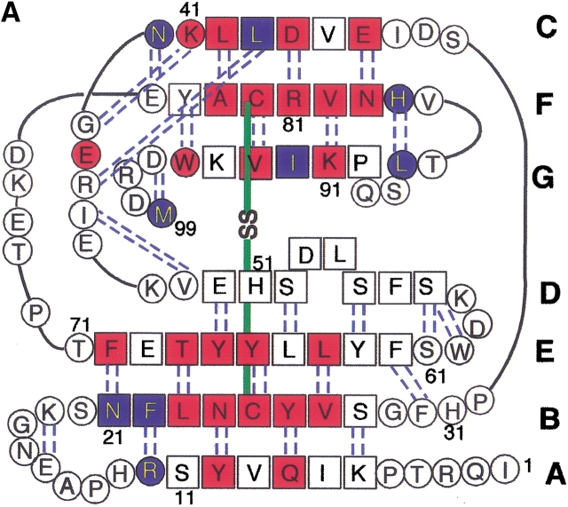
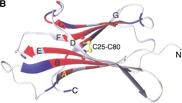
Fig. 1.
Amino acid sequence (A) and schematic structure (B) of β2-m. Secondary structures are indicated with hydrogen bonds (A) and the numbering of β-strands (A,B). The locations of strongly (red) and weakly (blue) protected amide protons are indicated. The figure was produced using MOLMOL (Koradi et al. 1996) with the structure (PDB entry 3HLA) reported by Bjorkman et al. (1987).


