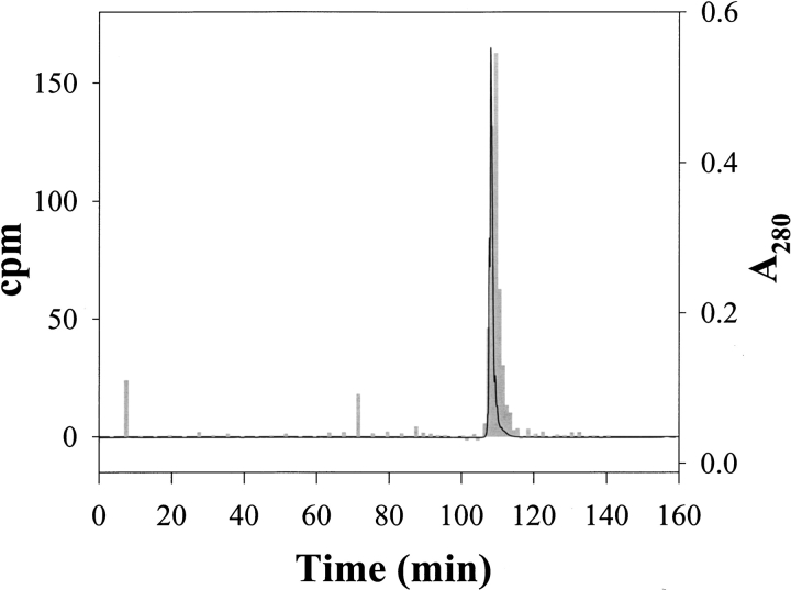
Fig. 3.
Separation of α-LA labeled with 3H-DZN by reversed-phase HPLC on a C4 column. The photolyzed sample contained 0.7 mM α-LA dissolved with 3.1 mM 3H-DZN (1 mCi/mmol) in 20 mM sodium phosphates buffer, pH 7.4. After the clean-up procedure described in Materials and Methods, the labeled protein sample (nearly 0.7 mg) was chromatographed through an HPLC C4 reversed-phase column (Vydac 214TP54, 4.6 mm × 250 mm) eluted isocratically with 0.05% aqueous TFA (20 min) followed by a linear gradient of acetonitrile:water (0 to 60% in 120 min) in 0.05% TFA at a 0.5 mL/min flow rate. α-LA eluted at 44% acetonitrile. Elution was monitored by both ultraviolet absorption at 280 nm (solid line) and by measurement of the radioactivity associated to each collected fraction (gray bars).

