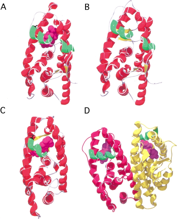
Fig. 1.

Representative structures of the ligand-binding domain of steroid and nuclear receptors. In (A–C), only the monomer is shown for simplicity; however, these structures are dimeric in the crystalline state. Helix 12, the N-terminal helix which caps the ligand-binding pocket, is indicated (green), and their respective ligands are shown in spacefilling (purple). (A) Ligand-binding domain of the progesterone receptor (PDB code 1A28; Williams and Sigler 1998) bound with progesterone. (B) Model of the ligand-binding domain of the glucocorticoid receptor; see Materials and Methods. (C) Ligand-binding domain of the retinoic acid receptor (RXR) (PDB code 2LBD; Renaud et al. 1995) bound to all-trans retinoic acid. (D) Dimeric form of the ligand-binding domain of the estrogen receptor in complex with diethylstilbestrol (PDB code 3ERD; Shiau et al. 1998).
