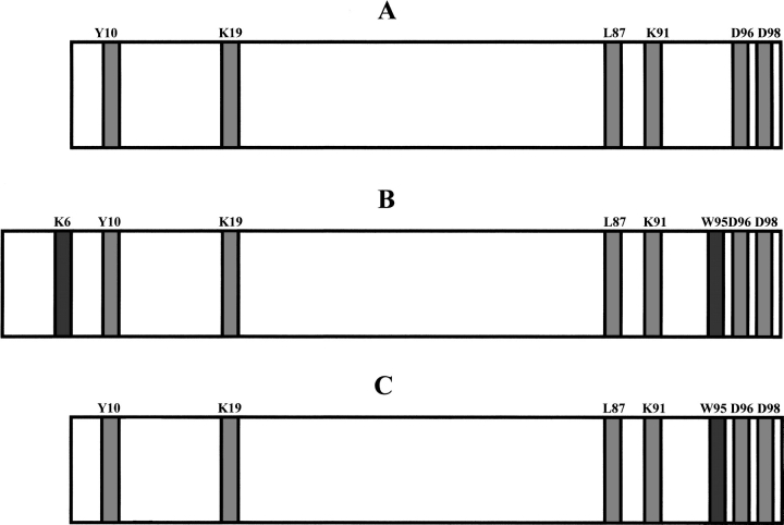
Fig. 5.
Schematic representation of the results obtained from limited proteolysis experiments. The preferential cleavage sites common to the soluble ΔN6β2-m (A) and to the fibrils originated from both intact β2-m (B) and ΔN6β2-m (C) are highlighted in light gray; those observed on intact β2-m (B) and ΔN6β2-m (C) at the fibrillar state only are highlighted in dark gray.

