Figure 2.
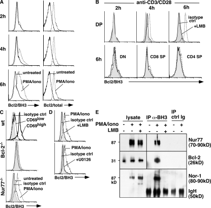
Bcl-2/BH3 exposure was observed in anti-CD3/CD28 and PMA/ionomycin-treated thymocytes. (A) Bcl-2/BH3 and Bcl-2 expression of DP cells from thymocytes cultured for 2, 4, and 6 h with 1.25 ng/ml PMA/0.5 μM ionomycin. Solid lines represent untreated thymocytes, and dotted lines represent treated thymocytes. (B) Bcl-2/BH3 exposure of DP cells from thymocytes cultured for 2, 4, and 6 h with 10 μg/ml anti-CD3/2 μg/ml anti-CD28 (plate-bound) and Bcl-2/BH3 exposure of DN and SP cells treated for 6 h. Here, the shaded area represents isotype control, dotted lines represent treated thymocytes, and solid lines represent treated thymocytes in the presence of leptomycin B. (C) Bcl-2/BH3 exposure of DP cells from Nur77−/− and Bcl-2−/− thymocytes cultured for 4 h with 2.5 ng/ml PMA/0.5 μM ionomycin. Shaded area represents isotype control, solid lines represent untreated thymocytes, and dotted lines represent treated thymocytes. Bcl-2/BH3 expression of CD69high and CD69low DP cells treated with 2.5 ng/ml PMA/0.5 μM ionomycin for 4 h is also shown. The shaded area represents isotype control, the solid line represents CD69low DP cells, and the thick line represents CD69high DP cells. (D) Pretreatment with 20 nM leptomycin B or 25 μM of the ERK5 inhibitor U0126 prevented exposure of the Bcl-2/BH3 domain in DP thymocytes stimulated for 4 h with PMA/ionomycin. Here, the shaded areas represent isotype control, dotted lines represent treated thymocytes, and solid lines represent PMA/ionomycin-treated thymocytes in the presence of leptomycin B or U0126. (E) PMA/ionomycin (PMA/iono) -stimulated thymocytes from lck-Bcl-2 mice were immunoprecipitated with anti-BH3 Bcl-2 antibodies or rabbit IgG, followed by blotting with antibodies specific for Nur77, Bcl-2, or Nor-1. Leptomycin (LMB) was added where indicated (+).

