Figure 1.
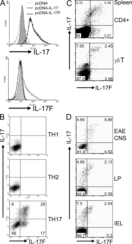
IL-17F is produced by IL-17–expressing T cells. (A) 293T cells transfected with pcDNA–IL-17 or pcDNA–IL-17F, or vector only, were fixed and stained with the indicated antibodies. (B) CD4+ T cells from OT-II mice were differentiated into Th1, Th2, and Th17 lineages. On day 5 of culture, CD4+ T cells were restimulated with PMA and ionomycin and stained with appropriate antibodies. (C) Splenocytes were activated with PMA and Ionomycin for 5 h, and IL-17– and IL-17F–expressing cells were assessed by intracellular staining on CD4+ or TCRγδ+ gates. (D) IL-17F and IL-17F–expressing cells in CNS infiltrates of mice with EAE, and in the lamina propria and intestinal intraepithelial lymphocytes were analyzed by intracellular staining with CD4+ gating. CNS infiltrates were isolated from perfused mice on day 12 after the second immunization. Data are representative of at least two independent experiments with similar results (percentages are shown).

