Figure 1.
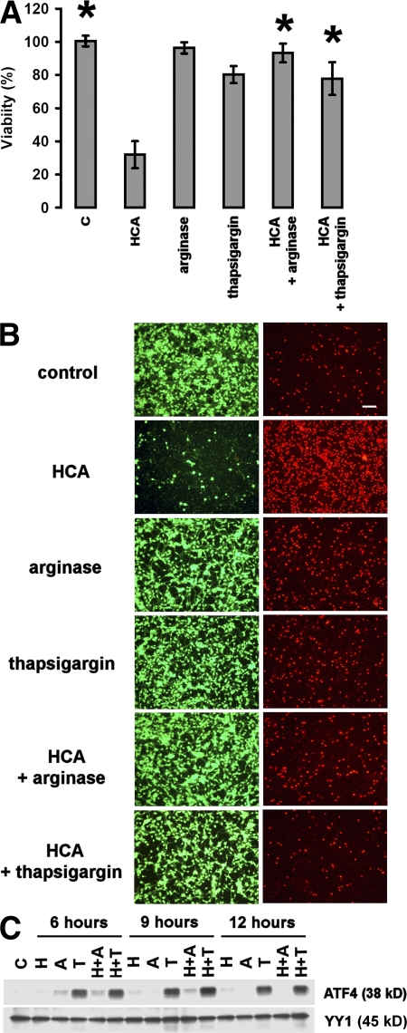
Amino acid depletion and thapsigargin treatment induce ATF4 and protect embryonic cortical neurons from oxidative stress–induced cell death. (A) Cortical neuronal cultures (1 d in vitro) were treated with a vehicle control (shown as C), 10 mM of the glutamate analogue HCA, 1 μg/ml arginase, 1 μM thapsigargin, 1 μg/ml arginase and 10 mM HCA, or 10 mM HCA and 1 μM thapsigargin. 24 h later, cell viability was determined using the MTT assay. The graph depicts mean (compared with control) ± SD calculated from three separate experiments for each group (n = 25). *, P < 0.05 from HCA-treated cultures by the Kruskal-Wallis test followed by Dunn's multiple comparisons test. (B) Live/dead assay of cortical neuronal cultures (2 d in vitro). Live cells were detected by uptake and trapping of calcein-AM (green fluorescence). Dead cells failed to trap calcein but were freely permeable to the highly charged DNA intercalating dye ethidium homodimer (red fluorescence). Bar, 50 μm. (C) Treatment with 10 mM HCA (shown as H) and 1 μg/ml arginase (shown as A) or 1 μM thapsigargin (shown as T), alone or in combination with HCA, increases the expression of ATF4 in cultured cortical neurons as compared with vehicle-treated control (shown as C). Cells were harvested at the indicated time points, and nuclear extracts were separated using gel electrophoresis and immunodetected using an antibody against ATF4. YY1 was monitored as a loading control. The immunoblot is a representative example of three experiments.

