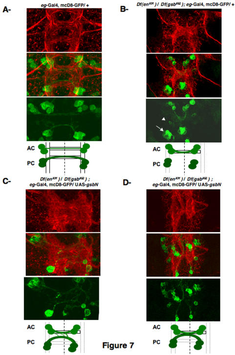
Figure 7. Behavior of eagle-positive neurons.
Flat preparations are shown of stage 15 eagle-Gal4, UAS-mcD8-GFP embryos, labeled with a Cy3-conjugated anti-HRP antibody to visualize the VNC architecture (red) upper panels; and with a polyclonal anti-GFP antibody, secondarily detected by Cy2-anti rabbit (green) lower panels, with the merged images in the middle. To help the lecture of the phenotypes, a diagram is provided. Only one segment is shown, but the corresponding entire cords are provided on Figure S1. eagle-positive neuronal behavior is shown A in a wild-type background, B in the transheterozygous (Df enX31/Df gsbX62) background (arrowhead indicates neuronal progeny of the NB 6-4, and arrow indicates neuronal progeny of the NB 7-3), C and D in the transheterozygous (Df enX31/Df gsbX62) background, when restoring GsbN expression in eagle-positive cells. Note that when commissures appear thicker, in 56% of the segments, two types of results are obtained and correspond to the pictures provided in C and D.

