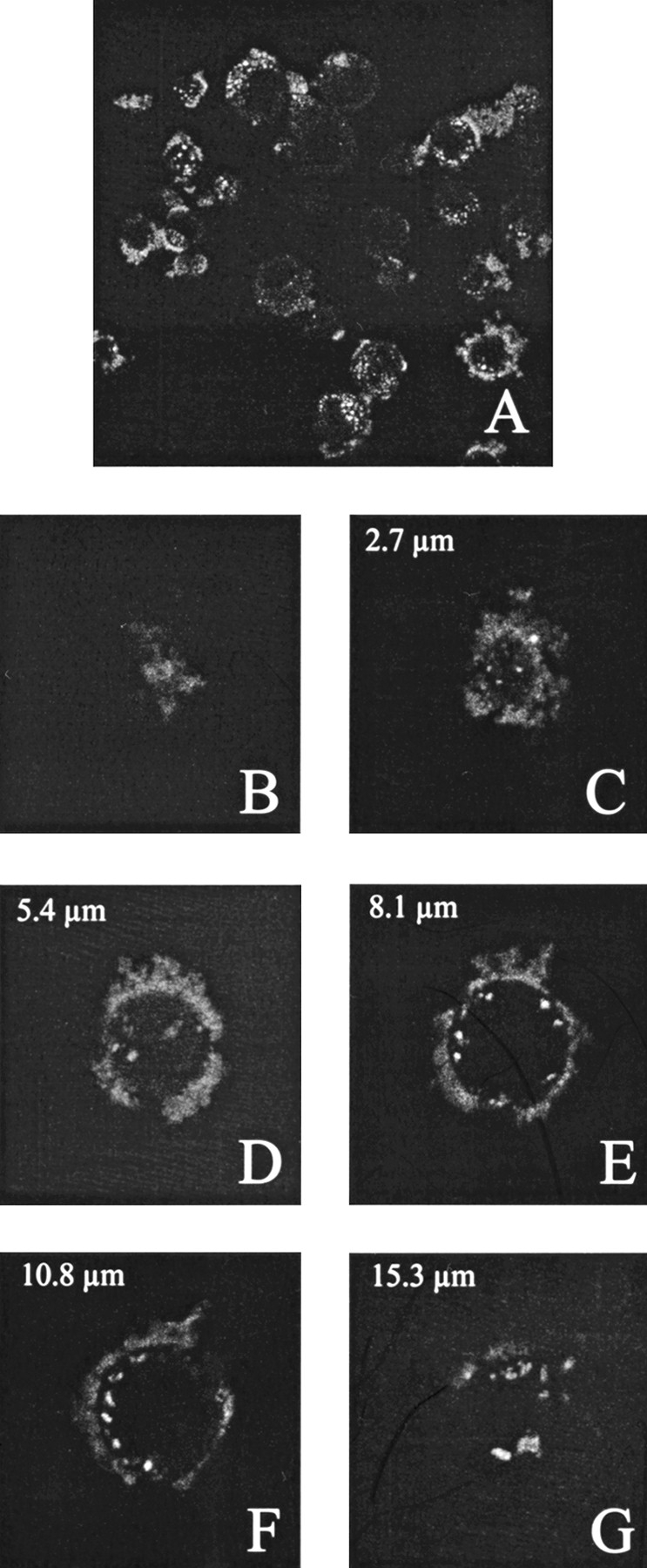
Fig. 10.

Cell type-specific targeting of antibody-presenting VLPs of VP1-Z. SK-BR-3 cells were grown to a confluency of ∼ 70%. Subsequently, the cells were incubated with VLPs of VP1-Z, which were decorated with the antibody Herceptin and packaged with fluorescence-labeled DNA. After overnight incubation nonbound VLP were removed by excessive washing and the cells were fixed using paraformaldehyde. (A) Overview of fluorescence-labeled cells. (B–G) Z series of a single cell. Fluorescence staining could be observed on the cell surface and inside the cells (D,E,F).
