Fig. 1.

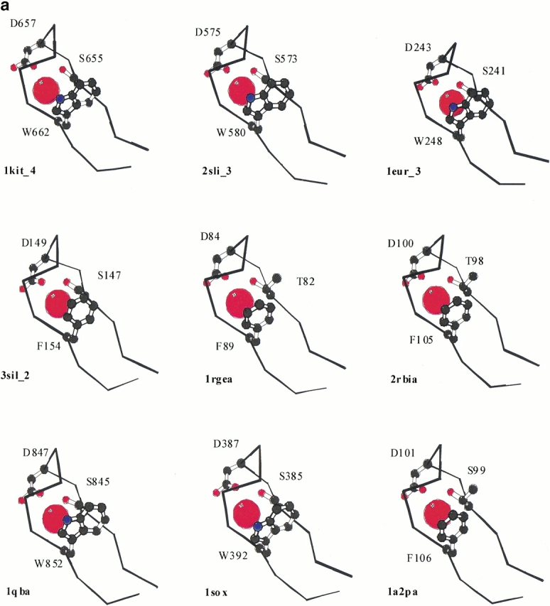
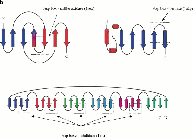
(a) Cα traces of Asp box structures shown in Table 1 represented using Molscript (Kraulis 1991). Only the best match from each of the β propellers is shown. PDB codes are as for Table 1. The side chain atoms of the core conserved residues are shown in ball and stick representation. The one letter amino acid codes and residue numbers are given. Water molecules found in equivalent locations in all structures are illustrated as red spheres. (b) Schematic representation of the location of Asp box motifs within different protein topologies. Amino and carboxy termini are labeled N and C, respectively. Arrows represent β strands, and cylinders α helices. Asp boxes identified in the structural search are boxed with dotted lines.

