
Fig. 1.
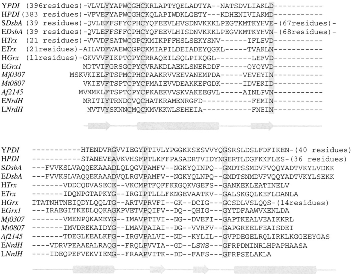
Sequence alignments of several protein disulfide oxidoreductases. The approximate locations of secondary structural elements in the minimal thioredoxin fold are displayed below the alignment. The alignment is based on highly conserved residues or residue types (filled boxes) as well as secondary structures based on prediction or from published crystal and NMR data. The identities of aligned proteins are as follows: S. cervisae PDI b domain (YPDI), H. sapiens PDI b domain (HPDI), S. typhimurium DsbA (SDsbA), E. coli DsbA (EDsbA), H. sapiens Trx (HTrx), E. coli Trx (ETrx), H. sapiens Grx (HGrx), E. coli Grx-1 (EGrx1), Mj0307, Mt0807, Af2145, E. coli NrdH (ENrdH), and L. lactis NrdH (LNrdH).