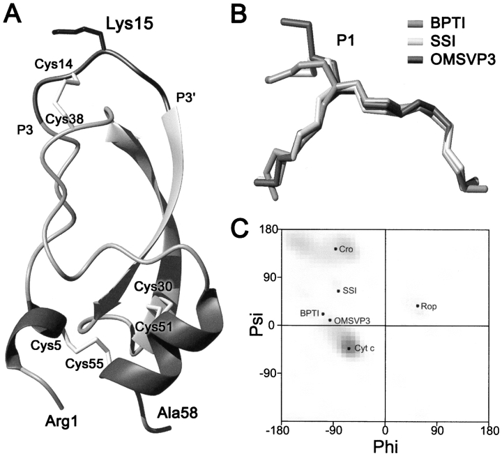
Fig. 1.
(A) Schematic representation of the crystal structure of BPTI (Wlodawer et al. 1987). Solvent-exposed Lys15 side chain is shown in the central part of the canonical-binding loop together with three disulfide bonds and secondary structure elements. (B) Superimposition of the main-chain conformation of the protease-binding loop (P3–P3′ segment) of BPTI (PDB code 5pti), SSI (3ssi), and OMTKY3 (2ovo). (C) The Ramachandran map showing ϕ, ψ angles adopted by the surface-exposed residues of several proteins that are mentoned in the text.

