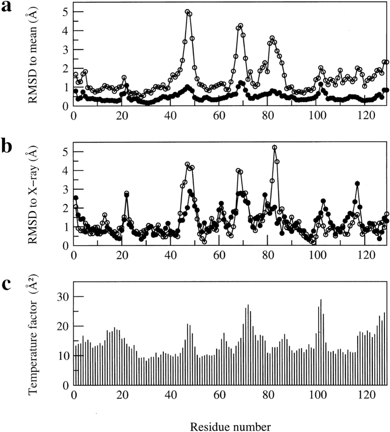
Fig. 1.
Comparison of (a) the average backbone RMSD values (in Å) from the mean structure and (b) the backbone RMSD values (in Å) between the mean structure and the X-ray structure (pdb code 193L) for the refined structures of hen lysozyme reported here and for those reported previously (Smith et al. 1993). In each case, the RMSD values are shown as a function of residue number, (filled circles) the ensemble of 50 refined structures in set 1 reported here, (open circles) the set of 16 structures reported previously (Smith et al. 1993). (c) shows the temperature factors (in Å2) of main-chain N atoms in the X-ray structure of Vaney et al. (1996) with pdb code 193L.

