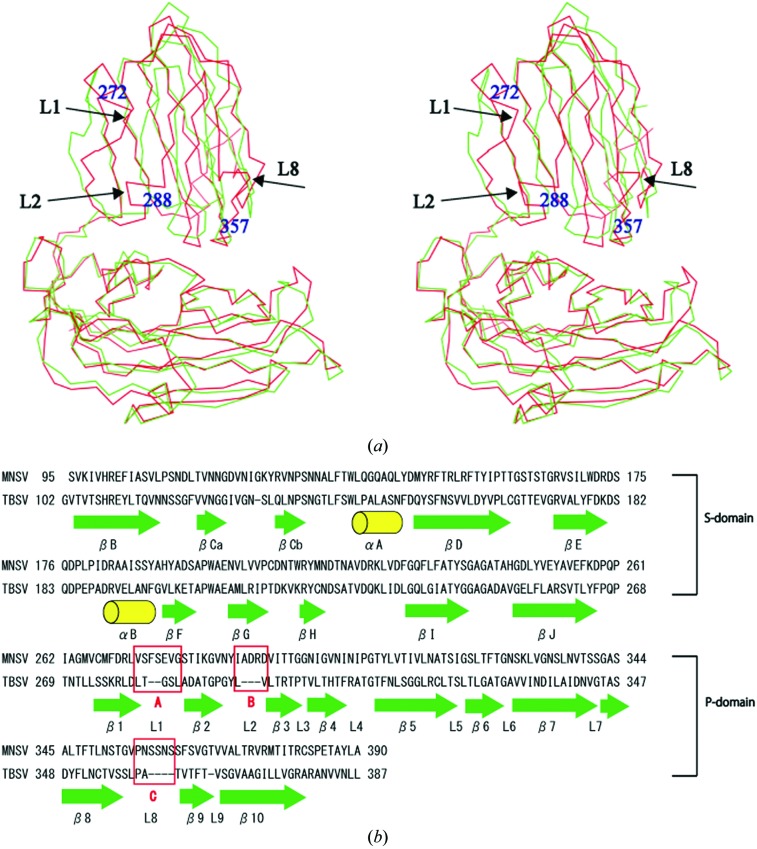
Figure 4.
Comparison of the coat-protein structures of MNSV and TBSV. (a) Shown here are stereo images of the A subunits of MNSV (red) and TBSV (green). Arrows represent insertion sequences. (b) Structure-based alignment of the coat proteins of MNSV and TBSV. Green arrows represent β-sheet structure and yellow cylinders represent α-helices. The red letters A (272–278), B (288–291) and C (357–362) represent insertions.

