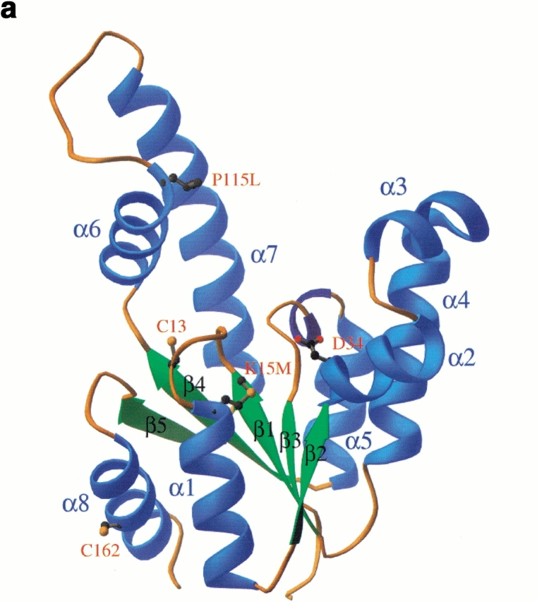
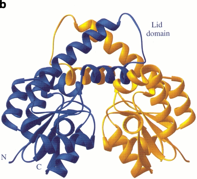
Fig. 4.

The three-dimensional structure of shikimate kinase mutant K15M. (a) A ribbon representation of the enzyme monomer colored according to secondary structure, with the position of residues mutated in this study shown in ball and stick. (b) Ribbon representation of the two independent molecules in the asymmetric unit. The extensive contacts of neighboring lid domains lead to a stabilization of this part of the molecule that is not visible in the native crystal structure. Both diagrams were generated using RIBBONS (Carson 1991).

