
Fig. 3.
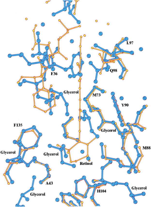
Superposition of the structures of rRBP and the retinol bound form of human retinol-binding protein (RBP; Cowan et al. 1990), showing the conformational change of the side-chain of Phe 36 and the binding of glycerol molecules in the place of retinol in the interior of the β-barrel. Recombinant RBP is depicted in blue; holo-RBP, in yellow.