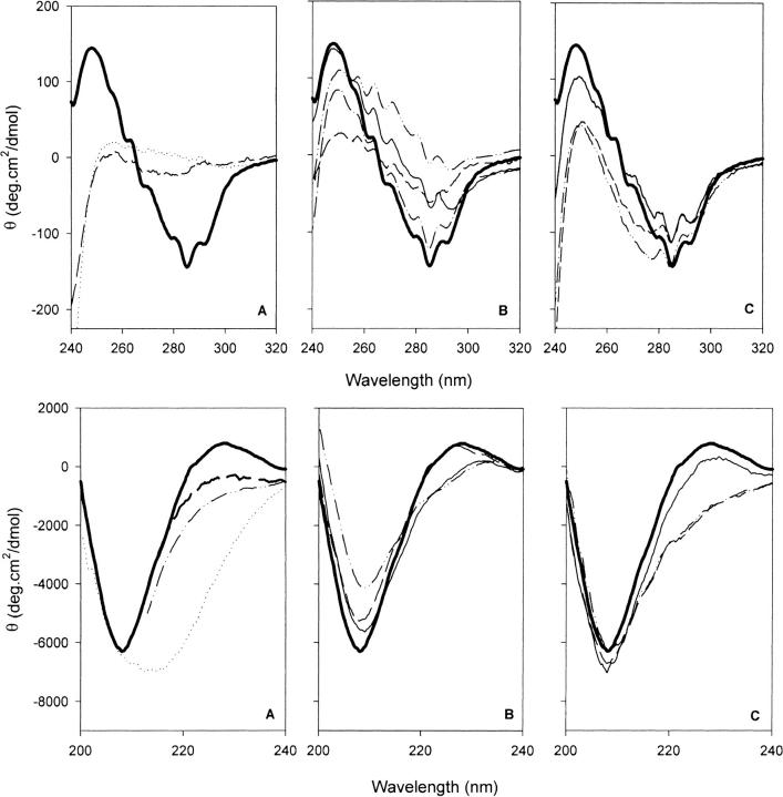
Fig. 5.
Near-ultraviolet (UV; 240 to 320 nm) and far-UV (200 to 240nm) CD spectra of recombinant retinol-binding protein (RBP) and variants. (A) Wild-type RBP: Native state at pH 7.4 and 20°C (thick solid line); unfolded state, 6 M GndHCl at pH 7.0 (broken line); and molten globule state at pH 2.0 (dotted line). (B) Mutants with substitutions for W67, W91, and W105: rRBP67L/91H/105F (– • •), rRBP67L/91H (– – –), rRBP91H (– •), and rRBP105F (dotted line). (C) Mutants with substitutions for G22, W24, and/or R139: rRBP24Y (broken line), rRBP24L (– • •), and rRBP139Q (thin solid line). The spectrum of native wild-type RBP is shown as a bold continuous line in all plots. Near-UV spectra were measured using a pathlength of 1 cm; far-UV spectra, with a pathlength of 0.1 cm.

