
Fig. 3.
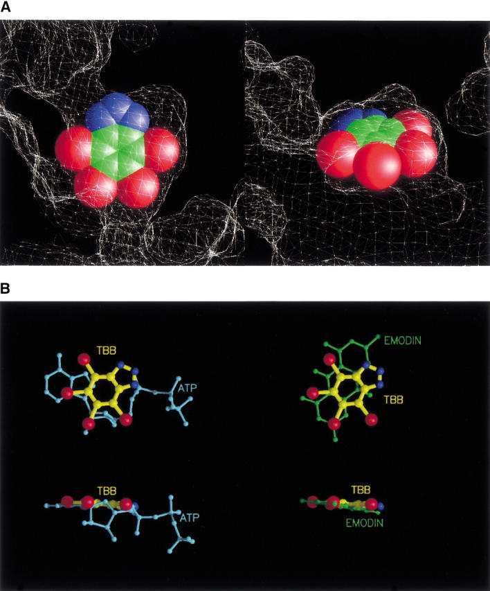
(A) Two clipped views of the active site showing the inhibitor (as cpk model) fitting the cavity are shown. The molecular surface of the protein is represented as a white mesh. Bromine atoms are in red, carbon atoms in green, and nitrogen atoms in blue. (B) Position of TBB with respect to that of ATP (cyan) and emodin (green) in the catalytic site is shown from different points of view. Inhibitor rings lay practically in the same plane of the purine moiety of the natural cosubstrates (bottom).