Fig. 4.

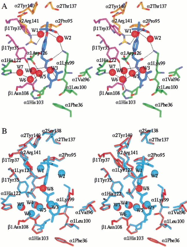
Stereoview of the allosteric site of effector 801. Similar effector environment is observed at the effector 802 site. Protein and effector atoms are shown in stick, and waters are spheres. For clarity, not all residues lining the allosteric binding site are shown. (A) Interactions between effector 801 (cyan) and Hb residues from the α1 subunit (green), α2 subunit (gold), and β1 subunit (magenta) are shown. Oxygen and nitrogen atoms are colored red and blue, respectively. Hydrogen bonds are indicated by black dashes. (B) Superimposition of the allosteric site of the native deoxy Hb structure (red) and the RSR-13:deoxy Hb complex structure (cyan). Note that the water molecules Wat 3, Wat 4, Wat 5, and Wat 7 are conserved, while Wat 1, Wat 2, and Wat 6 are unique to the complexed structure, and Wat 8 is unique to the native structure. The figures were generated using InsightII (Molecular Simulations Inc.) and labeled with Showcase.
