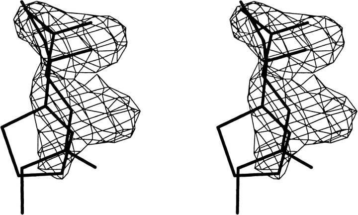
Fig. 5.
A stereo diagram shows the structures of SBT and HMH superimposed on electron density present in the active site of MUP-I isolated from liver following a 5000 K simulated annealing refinement in the absence of ligand. The HMH ketone oxygen and SBT nitrogen occupy similar positions in the ligand binding site. The Fo-Fc map is contoured at 3.0 σ. The closed ring form of HMH (Fig. 1 ▶) did not fit the electron density (data not shown).

