
Fig. 5.
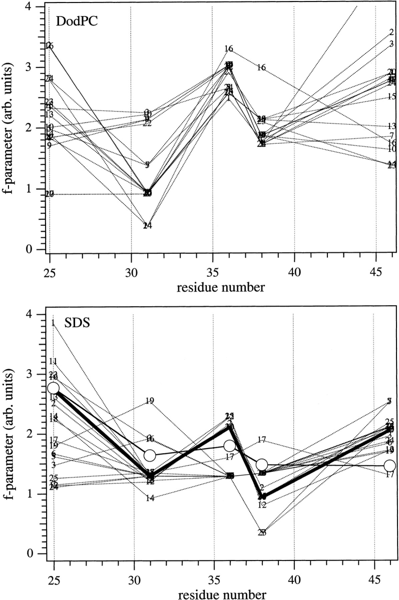
Sequence profiles for the f-parameter of M13 coat protein structures with single cysteine–maleimide replacements, in the absence of a lipid bilayer. Each x-coordinate represents a single mutant of the original families of structures determined in DodPC (top) or in SDS (bottom), in which the cysteine–maleimide side chain was locally optimized after amino acid replacement. Structure numbers indicated in the figure are those in the PDB files 2CPB and 2CPS for NMR-based structures in DodPC and SDS micelles, respectively, determined by Papavoine et al. (1998). (Bottom) The heavy solid line indicates the selected structure no. 20; open circles are the experimental EPR outer hyperfine splittings scaled to best match this profile.