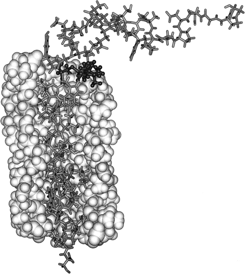
Fig. 6.

Structure of Cys 25-spin labeled M13 coat protein (stick representation) surrounded by a single bilayer solvation shell of DOPC phospholipids. For clarity, only part of the lipid shell is shown—in space-filling representation. The structure was chosen to satisfy the experimental EPR constraints and was obtained by geometry optimization of the lipids and all protein side chains. Cys 25, with the 5-maleimidoproxyl label bound to it, is enhanced with dark ball-and-stick representation. The figure is created using Insight II.
