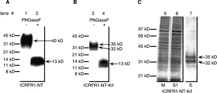
Fig. 2.
Polyacrylamide gel analysis of rCRFR1-NT and rCRFR1-NT-Kif. For deglycosylation with PNGaseF, approximately 100 ng Ni-affinity-purified rCRFR1-NT (A) or 37.5 μL of medium containing rCRFR1-NT-Kif (B) were applied to SDS-PAGE followed by Western blot and immunodetection. The absence or presence of PNGaseF is indicated. (C) SDS-PAGE of affinity-purified rCRFR1-NT-Kif was performed by application of 37.5 μL medium of transfected HEK 293 cells (M), 37.5 μL supernatant after adsorption on Ni-affinity resin (S1), and 30 μL of the third elution fraction (E). Proteins were detected by silver staining.

