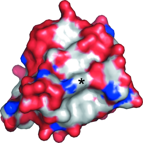
Figure 2.
Surface representation of the endophilin SH3 domain. The surface is color-coded by element, with red representing oxygen, blue nitrogen and gray carbon. The ligand-binding groove runs approximately vertically in this view. The base of the groove can be seen as the gray (hydrophobic) stripe flanked by red ‘walls’ on either side. The side chain of Trp343 (indicated by an asterisk) forms a ridge across the base of the groove. Rotating the molecule shown in this figure by approximately 35° about a vertical axis will result in a view similar to that shown in Fig. 1 ▶.

