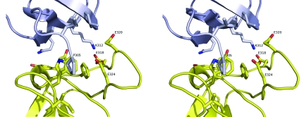
Figure 4.
Stereoview of the crystal-packing interaction involving the ligand-binding site. This figure corresponds to an expanded view of the region highlighted in the gray box in Fig. 3 ▶. Residue Pro305 at the N-terminus of the gray molecule (foreground) packs against Trp343 in the ligand-binding groove of the yellow molecule and Lys312 of the gray molecule inserts into an acidic pocket formed by three glutamate residues on the yellow molecule.

