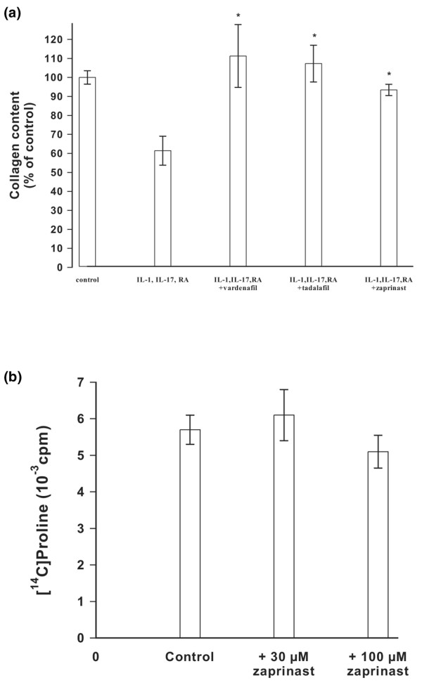
Figure 3.

Quantitative analysis of collagen degradation and its inhibition. (a) Cartilage explants were incubated with IL-1α, IL-17 and retinoic acid in the absence or presence of zaprinast, tadalafil, or vardenafil at concentrations of 50 μmol/l for 4 weeks at 37°C. The amount of chymotrypsin-resistant collagen was determined as hydroxyproline. The values were related to the control that contained 23 μg hydroxyproline/mg cartilage as 100%. The bars indicate the standard deviation of four determinations; *P < 0.05. (b) Bovine chondrocytes were grown in alginate beads and and collagen was labeled by incorporation of [14C]proline in the absence or presence of 30 μmol/l and 100 μmol/l zaprinast. The amount of [14C]collagen within the alginate beads was determined after 24 hours. The bars indicate the standard deviation of four determinations.
