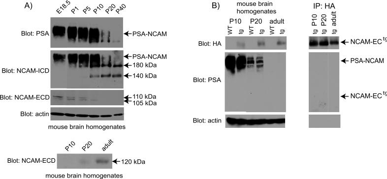
Figure 1. Expression of different NCAM isoforms and cleavage products during development.
A) NCAM, PSA-NCAM and cleavage fragments during development. Brain homogenates (75 μg) from E18.5 to P40 mice were separated by SDS-PAGE, and blotted using antibodies to PSA (top panel), NCAM-ICD (clone OB11; second panel), and NCAM-ECD (clone H300; third panel). Arrows indicate the size of PSA-NCAM, NCAM180, NCAM140, and cleavage fragments representing the NCAM extracellular domain (105, 110 kDa). NCAM120 expression (bottom panel). Homogenates were blotted using the NCAM-ECD antibody. Actin blots (fourth panel) were used as a loading control. Exposure times were: 1 min (NCAM-ICD, actin), 5 min (PSA, NCAM-ECD bottom panel), 1 h (NCAM-ECD third panel). B) NCAM-ECtg expression and polysialylation. The left set of panels depicts Western blots of mouse brain lysates. For PSA detection, NCAM-ECtg was immunoprecipitated using HA antibodies prior to SDS-PAGE and blots are depicted in the right set of panels. Western blots were performed using antibodies to HA (top panels, 1 min exposure), PSA (middle panels, 5 min exposure), and actin (loading control, bottom panels, 1 min exposure).

