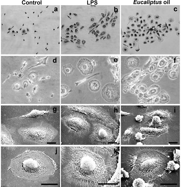
Figure 1.
Morphological features of human MDMs after 24 h of in vitro treatment with Eucalyptus oil. a-f, phase-contrast microscopy after Wright Giemsa staining: a, d, untreated control; b, e, MDMs stimulated with 0.1 μg/ml of bacterial lipopolysaccharide (LPS); c, f, MDMs treated with 0.016% Eucalyptus oil; a, b, c, original magnification: 20×;d, e, f, original magnification: 40×. g-l, Scanning electron microscopy of untreated (g, j), LPS treated (h, k) and Eucalyptus oil treated MDMs (i, l). Bars: 20 μm.

