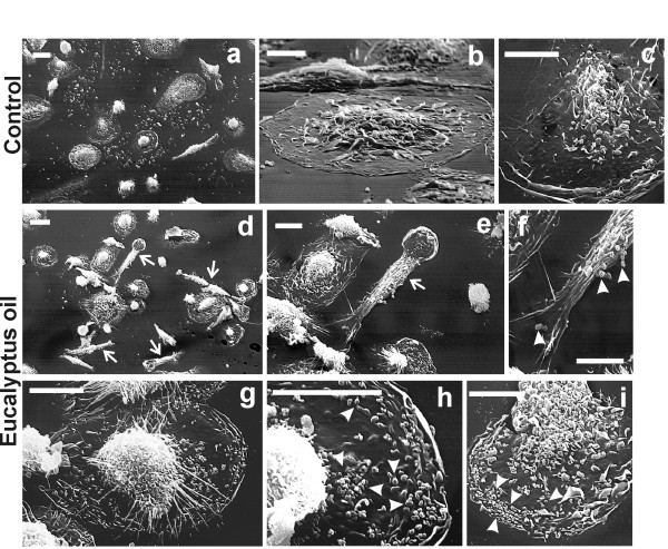
Figure 3.
Morphological features of 24 h EO treated human MDMs after in vitro administration of polystyrene beads. Scanning electron microscopy of untreated (a-c) and 0.008% Eucalyptus oil treated MDMs (d-i) showing the presence, in EO treated cultures, of numerous polarised cells exhibiting elongated lamellopodia and filopodia (arrows in panels d and e), indicative of a pseudopodial activity. Arrowheads in panels f, h, i point to phagocytosed beads, more numerous respect to the untreated control (c). Bars: a, d = 20 μm; remaining panels = 10 μm.

