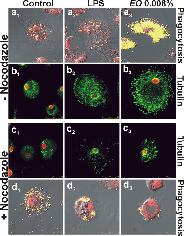
Figure 5.
Effect of microtubule destabilization by nocodazole on phagocytic activity of Eucalyptus oil stimulated human MDMs. a, d, Confocal microscopy images, showing the beads uptake (yellow hue) in control (a1, d1), LPS pre-treated (a2, d2) and EO pre-treated MDMs (a3, d3), in absence (a) or in presence (d) of nocodazole treatment; cells were counter-stained with PI (red hue). Merged images with differential interference contrast, used to visualize cell morphology, are shown. b, c, Confocal microscopy images, showing the microtubular network (green hue) in nocodazole treated and untreated cells: b1, c1, controls; b2, c2, LPS pre-treated MDMs; b3, c3, EO pre-treated MDMs.

