
Fig. 11.
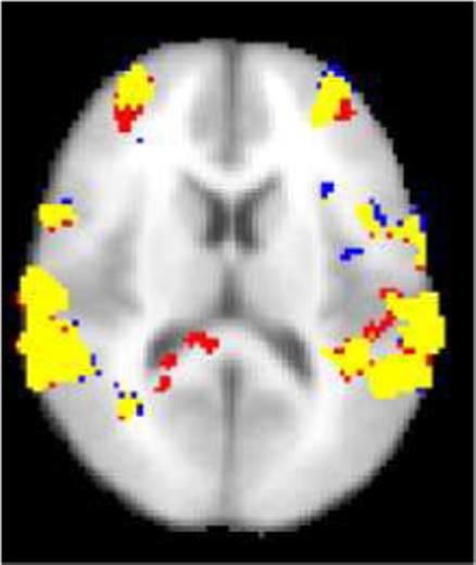
An axial slice of the map showing regions of the greater dual task activity than single task activity in the group analysis (significance threshold P = 0.05, whole-brain corrected using Gaussian Random Field Theory). Yellow: The voxel is activated both for the denoised dataset and the original dataset. Red: The voxel is activated only for the original dataset. Blue: The voxel is activated only for the denoised dataset. The main effect of denoising was the reduction of the activations detected in white matter posterior to the ventricles. The NP threshold was 0.05.