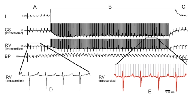
Figure 2: A typical episode of a stun gun shock across the abdomen (nonthoracic vector) that does not result in stimulation of the myocardium. The surface electrocardiogram lead 1, intracardiac electrograms from the coronary sinus, the right ventricle apex and blood pressure in the descending aorta are shown. Panel A illustrates the regular rhythm before the discharge, which is very similar to the rhythm and rate in panel C. The intracardiac electrograms, as illustrated in panels D and E, do not show any significant change in rate morphology and are not phase-locked (no temporal relation between stimuli and the electrogram) with the stun gun discharge. Note also the lack of perturbation of blood pressure during the discharge. Reproduced with permission from Elsevier (Nanthakumar et al24). Note: CS = coronary sinus, RV = right ventricular, BP = blood pressure.
