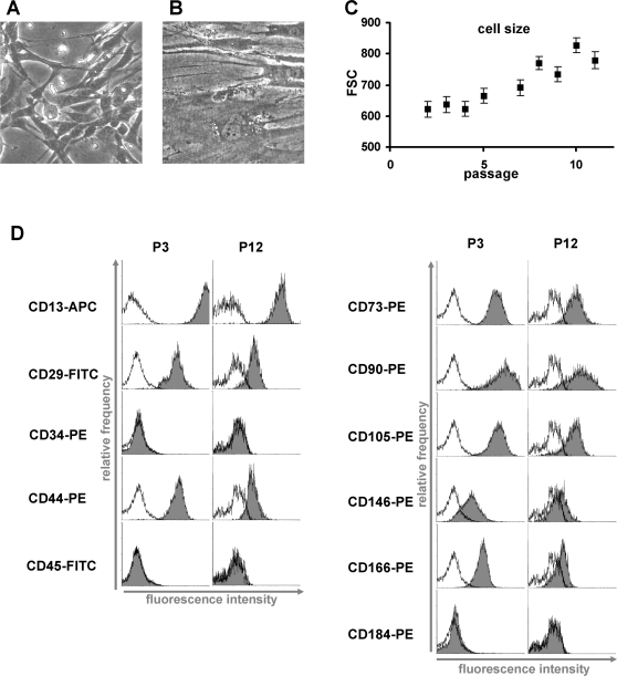
Figure 2. Morphologic changes and immunophenotype upon senescence.
Replicative senescence is reflected by dramatic changes in morphology. Cells enlarge, generate more vacuoles and cellular debris and ultimately stop proliferation. Representative morphology of MSC in early (P3) and senescent passage (P12) is presented (A, B). The continuous increase in cell size and granularity is reflected by the increasing forward-scatter signal in flow cytometry (FSC, ±SD; C). Immunophenotypic analysis of all MSC preparations was in accordance with the literature whereby the detection level for positive markers was much higher in early passages compared to late passages (black line = autofluorescence; D). A representative analysis of three preparations is demonstrated.

