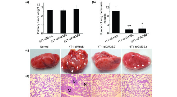
Figure 5.
Tumor formation and lung metastases 30 days after tumor implantation. (a) Primary tumor weights. (b) The average numbers of lung metastatic nodules. (c) Representative photos of the lungs. The arrows point to the metastatic nodules in lung. (d) Representative hematoxylin and eosin staining sections of the lungs were photographed at 40× magnification. *P < 0.05. M, metastatic nodule; N, normal.

