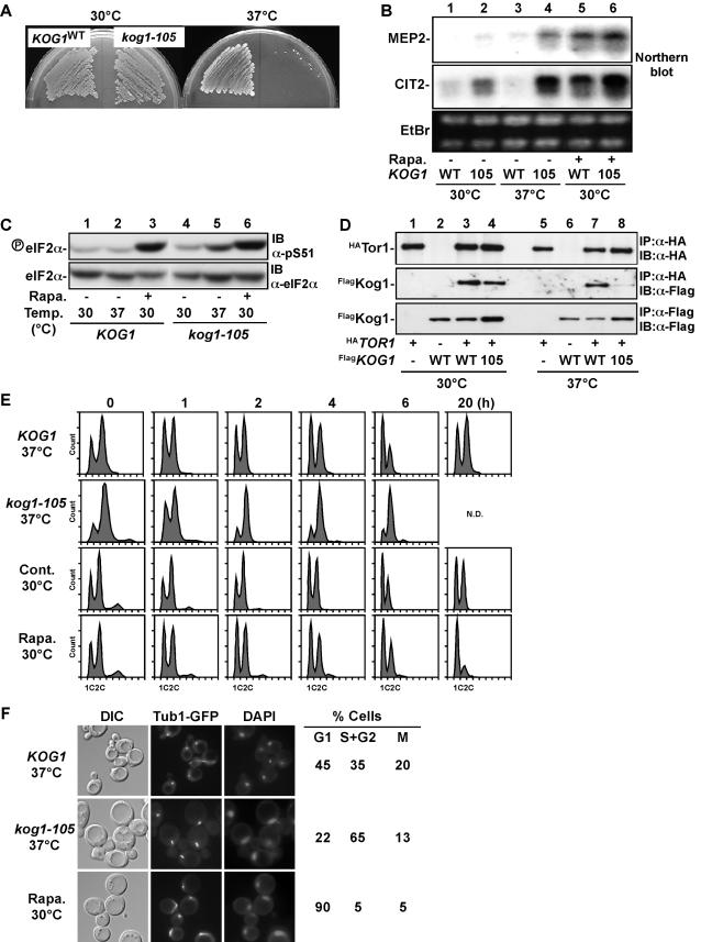
Figure 1. Characterization of kog1-105, a temperature-sensitive mutant of KOG1 gene.
(A) Wild-type (KOG1, YYK409) and kog1-105 (YYK410) were grown on YEPD at 30°C or 37°C for 2 days. (B) Enhanced expression of starvation-induced gene in kog1-105. Cells grown at 30°C were incubated at 37°C or 30°C in the presence of 200 ng/ml of rapamycin (Rapa.) for 2 h. Total RNA was extracted and analyzed for expression of MEP2 (top panel) and CIT2 (middle panel) genes by northern blot. RNA amount was monitored by ethidium bromide staining (bottom panel). (C) Phosphorylation of eIF-2α is enhanced in kog1-105. Cells grown at 30°C were incubated as described in (b). Cell lysate was analyzed by immunoblot using an anti-phospho eIF-2α antibody (pS51, top panel) and an anti-eIF-2α peptide antibody (bottom panel). (D) Tor1-Kog1 association is unstable in kog1-105. HA-tagged Tor1 (HATor1) was immunoprecipitated from wild-type (YAN86) and kog1-105 cells (YAN103) (top panel), and co-precipitated Flag-tagged Kog1 (FlagKog1) was detected by immunoblot (middle panel). Noted that protein amount of FlagKog1 was similar in wild-type and kog1-105 (bottom panel). (E) Wild-type (KOG1 (YYK409)) and kog1-105 (YYK410) cells grown on YEPD at 30°C were incubated at 37°C or 30°C in the presence of 200 ng/ml of rapamycin for the indicated times. DNA content was determined by FACS analysis. N.D.; not determined because of loss of viability. (F) Wild-type (KOG1 (KLY4206)) and kog1-105 (YYK834) cells expressing GFP-tagged tubulin (Tub1-GFP) were treated as in (e) for 4 h. Spindle formation and DNA (DAPI staining) were observed by immunofluorescent microscope. Percentage of G1 (unbudded cell), S+G2 (budded cell with short spindle), and M phase (large budded cell with long spindle) cells was also determined.

