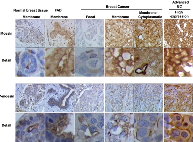
Figure 12. Moesin and P-moesin expression and sub-cellular localization in human normal breast tissue, benign fibroadenomas and breast cancers.
Histological sections (4 µm) from normal mammary glands, fibroadenomas or ER positive (ER+) breast cancers were used to identify the expression and localization of moesin and P-moesin with immunochemistry. Wild-type moesin as well as Thr558-phosphorylated are shown as brown labeling. The figure displays sample images from the tumors analyzed in Table 1. Particularly, The normal breast tissue comes from Patient 2, the fibroadenomas is from patient 3. The breast cancer showing focal staining is that of patient 20, the cancer with membrane staining comes from patient 13. The cancer showing mixed membrane/cytoplasmic staining is from patient 17. The “high expression” cancer is from patient 12.

