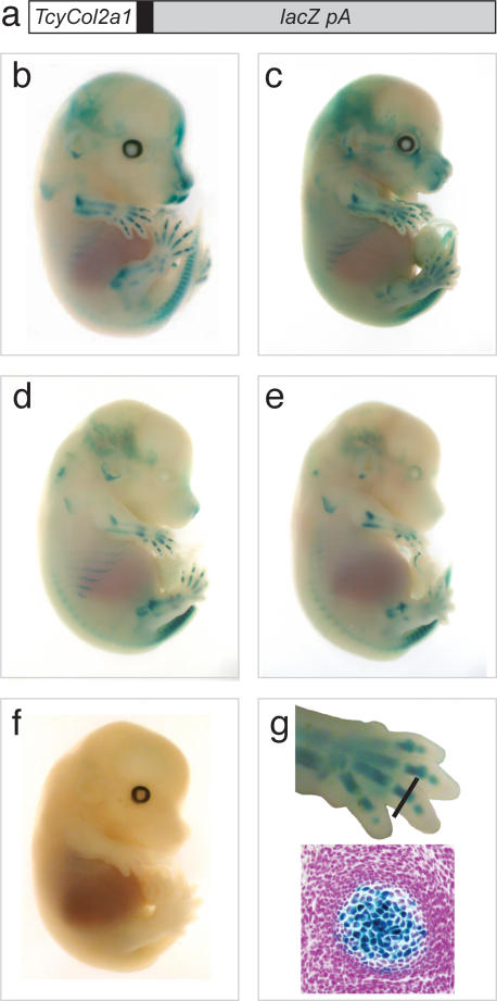
Figure 3. From extinction to gene expression.
Functional analysis of the thylacine non-coding DNA fragment. (a) Diagram of transgene construct. 4 copies of a 264-bp fragment containing the Thylacine Col2a1 enhancer (TcyCol2a1) region was ligated to the human b-globin minimal promoter (black box) and ligated to lacZpA. (b–e) X-gal stained 14.5 dpc TcyCol2a1-lacZpA transgenic mouse embryo showing varying levels of reporter gene expression within the developing cartilage (blue). (f) Non-transgenic littermate, negative control fetus. (g) Top panel; Magnified image of forelimb from fetus in (b) black line indicates the plane of section shown in (g) bottom panel. Bottom panel; Histological section of transgenic forelimb digit, showing lacZ-expressing chondrogenic tissue (blue) counterstained with eosin (pink).

