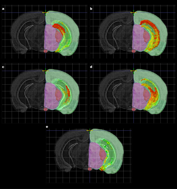
Figure 6.
Top 10 results for a set of spatial homology searches are shown overlaid for subregions of the hippocampus. The seeds used are shown in Figure 5: Prox1 for the dentate gyrus (a), Ptpru for CA1 (b), Cacng5 for CA2 (c), Prss35 for CA3 (d) and Cd44 for the ventral CA3 (e). The view is restricted to the hippocampal region. A rostral clipping plane and coronal atlas plate were set to isolate the middle of the hippocampal region. The 3D projections can be compared with the hippocampal structures shown in the Nissl-stained reference atlas on the left hand side of the figures.

