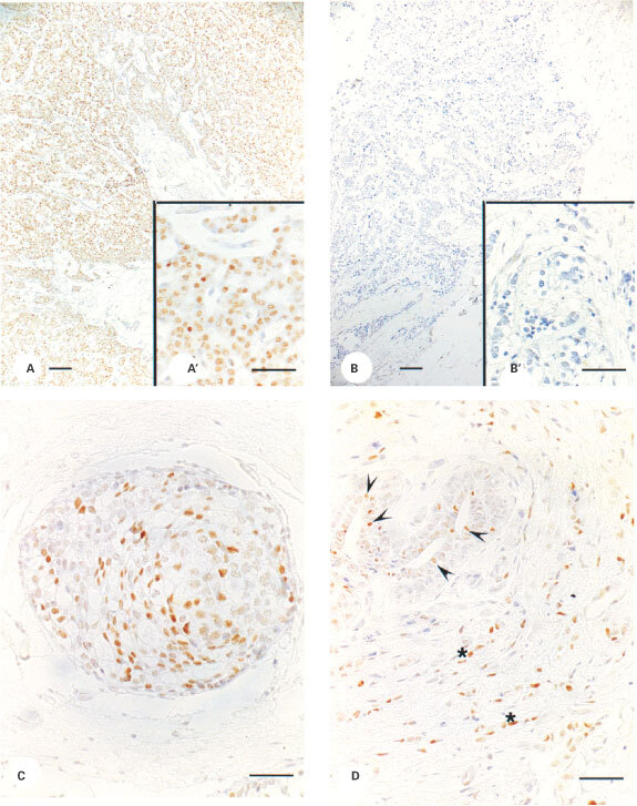
Figure 4.

Immunoexpression of ERβ in human breast tissues. Nuclear expression of ERβ protein was detected in 94% of the samples examined. (A,B) show examples of immunopositive (A, code 5580) and immunonegative (B, code 5667) staining of malignant tissue. Expression of ERβ was also noted in non-invasive ductal cancer (C) and in epithelial (D, arrowheads) and stromal (D, asterisks) cells in areas of breast not associated with malignant growth. (A,B), Magnification ×10, bar=100 μM, insets A′ and B′ are from the same tissues as A and B, magnification ×40, bar=50 μM. (C, D) Magnification ×40, bar=50 μM.
