
Figure 4.
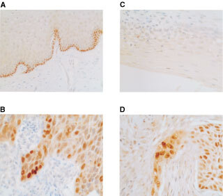
Immunocytochemical analysis of p73 and p14ARF in cervical epithelium. Sections were prepared from formalin-fixed, parrafin- embedded tissue samples as described in Materials and Methods. (A) Expression of p73 is in the basal layer of normal cervical epithelium. RT–PCR analysis revealed that this was full-length, p73α in all cases studied. (B) p73 protein is over-expressed in cervical SCC. RT–PCR analysis revealed expression of both full-length and p73 Δ2 in the majority of cases. (C) p14ARF protein expression is not detectable in normal cervical epithelium. (D) Over-expression of p14ARF in cervical SCC. Note the prominent nucleolar expression and sparing of normal tissue.