
Figure 4.
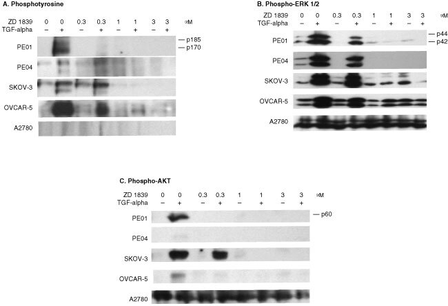
Western blot of ZD 1839 treated cell lines using (A) the anti-phosphotyrosine PY20 antibody, (B) the phospho-ERK NEB 9101 antibody and (C) the phospho-AKTS473 NEB 9271 antibody. Blots are shown for the PE01, PE04, SKOV-3, OVCAR-5 and A2780 cell lines. ZD 1839 was added 30 min prior to TGFα (1 nM) addition and lysates were collected after 15 min exposure to TGFα. Amounts of protein loaded were 75 μg lane−1 for the phosphotyrosine gel and 35 μg for the phospho-ERK and phospho-AKT gels. Exposure times varied from 20 s to 2 min but were the same for each antibody.