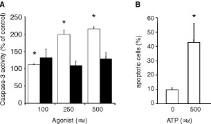
Figure 2.

Apoptotic effects of extracellular nucleotides. (A) ATP (white columns) but not ATPγS (black columns) dose-dependently induced caspase-3 activity in Kyse-140 cells after 48 h of incubation. Caspase-3 activity was measured as the cleavage of the fluorogenic substrate DEVD-AM. Means±s.e.m. of four independent experiments are given as the percentage of fluorescence compared to untreated control. (B) Nucleic DNA fragmentation of human oesophageal primary culture cells was determined using TUNEL assay. Incubation with 500 μM ATP for 48 h induced a significant increase of the proportion of nucleic fluorescence staining. The proportions of positively stained cells are shown as means±s.e.m. of three independent preparations.
