
Figure 2.
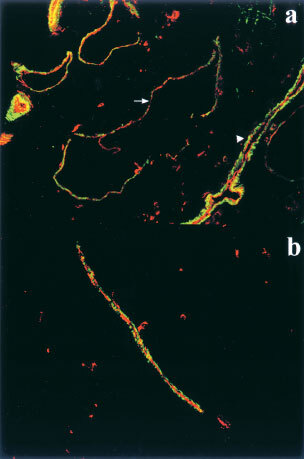
Ang1 prevents dissociation of smooth muscle cells from tumour edge-associated vessels. Confocal microscopic images of tumour edge-associated vessels were obtained by double immuno-fluorescent staining. Original magnification x200. Endothelial cells (red) were immunostained for CD31 and visualized with TRITC. Smooth muscle cells (green) were immunostained for α-actin and visualized with Green-FITC. (a) Many peripheral blood vessels in the control MCF-7 tumours are highly dilated and poorly supported by smooth muscle (arrow). Some blood vessels appear to have a normal smooth muscle layer (arrow head). (b) A typical edge-associated vessel in Ang1-overexpressing tumours showing an undilated lumen with good smooth muscle support.