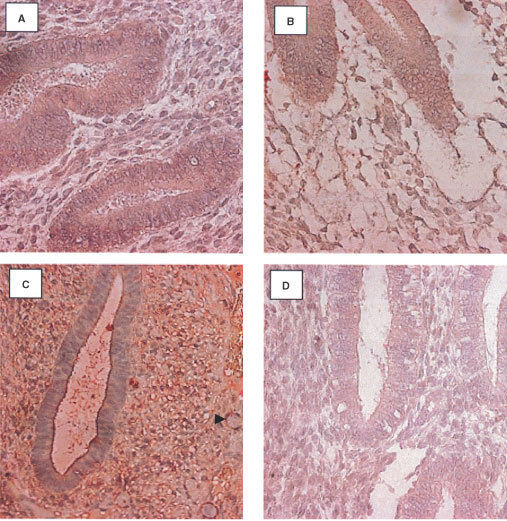
Figure 1.

Localization of aFGF in the endometrium using a streptABC alkaline phosphatase method. Positive staining appears red. (A) Weak expression in the normal endometrium (early secretory), (B) weak aFGF expression in the epithelium and stroma of the post menopausal endometrium, (C) increased expression in the epithelium and stroma of the premenopausal tamoxifen samples (early proliferative). (D) weak expression in the epithelium and stroma of the post menopausal tamoxifen exposed endometrium (Magnification ×400) (Arrowhead indicates position of a blood vessel).
