
Figure 3.
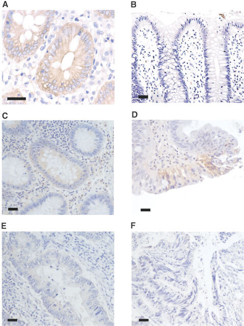
Immunohistochemical detection of MCT1 in normal, adenoma and carcinoma human colonic sections. Immunohistochemistry was carried out on normal, adenoma and carcinoma human colonic tissue using a horseradish peroxidase-conjugated secondary antibody as described in the Materials and Methods section. MCT1 staining is seen as a brown colouring contrasting with the blue/purple counterstained nuclei. (A) Healthy human colon (×400); (B) healthy human colon with MCT1 antibody pre-incubated with immunising peptide (×200); (C) tubular adenoma (×200); (D) tubular-villous adenoma (×200); (E) well differentiated carcinoma (×200); (F) poorly differentiated carcinoma (×200). Bars represent 50 μm. This data is summarised in Table 1.