
Figure 5.
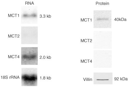
Assessment of the expression of MCT2 and MCT 4 in colon carcinoma measured by Western and Northern analyses. Post-nuclear membranes and total RNA were isolated from malignant colonic tissues, and analysed for MCT1, MCT2, MCT4, villin or 18S rRNA expression as described in the Methods section. Left panel: representative Northern blot showing abundance of MCT1 and MCT4 RNA in malignant colon. Nylon membranes were stripped and re-probed for 18S rRNA to confirm equality of loading. Right panel: representative Western blot showing the levels of MCT1 MCT2, and MCT4 proteins in malignant colon carcinoma tissues. Nitrocellulose membranes were stripped and re-probed for villin to confirm equality of protein loading. Note: specific immunoreactive bands were detected in control samples for MCT2 (in rat liver homogenate) and MCT4 (in rat heart tissue homogenate).