
Figure 6.
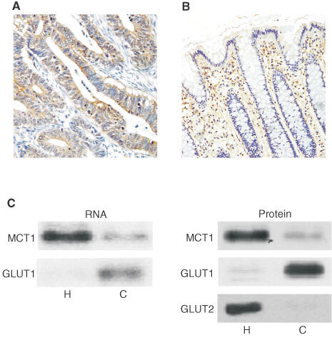
Abundance of GLUT1 in healthy and diseased colon. Immunohistochemistry was carried out on healthy and malignant human colonic tissue sections using a horseradish peroxidase conjugated secondary antibody as described under ‘Methods’. GLUT1 staining is seen as a brown colour contrasting with the blue/purple counterstained nuclei. (A) moderately differentiated carcinoma (×200). Bars represent 50 μm. Pre-incubation of antibody with immunising peptide resulted in absence of staining (data not shown). (B) Healthy colon (×200). Significant erythrocyte staining can be seen in the lamina propria. (C) Post-nuclear membranes and total RNA were extracted from healthy (H) and carcinoma (C) tissues and analysed for MCT1, GLUT1 and GLUT2 mRNA and protein abundance as described in ‘Methods’. Left panel: representative Northern blot indicating levels of MCT1 and GLUT1 mRNA (n=6). Right panel: Representative Western blot demonstrating levels of MCT1, GLUT1 and GLUT2 proteins (n=6).