
Figure 4.
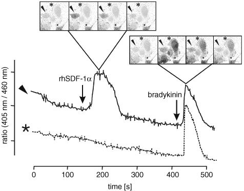
Ca2+ flux in A-498 cells in response to rhCXCL12α. Using confocal microscopy, we detected a heterogeneous reaction pattern of A-498 cells in response to rhCXCL12α stimuli. While stimulation with 2 μg ml−1 rhCXCL12α caused a rapid and robust increase of intracellular calcium in a fraction of A-498 cells (e.g. cell marked with an arrowhead), others did not respond to the stimulus (e.g. cell marked with a star). In contrast, a large increase in transient calcium was evoked by stimulation with 100 nmol bradykinin in all A-498 cells. Upper part of Figure=microscopy of single cells; lower part of Figure=intracellular Ca2+ flux visualised by fura-2 staining; arrow head, …rhCXCL12α responding A-498cell; *-rhCXCL12α non responding A-498 cell.